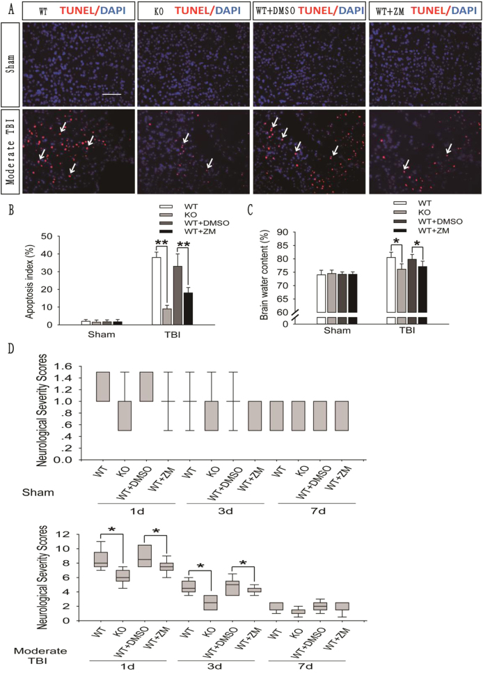
Fig. 5. A2AR KO or inhibition improved the prognosis of brain injury after moderate TBI.
a Images of apoptotic cells in cortical brain sections obtained from the WT + TBI, A2AR KO + TBI and WT + TBI + ZM241385 groups using TUNEL staining 1 day after sham surgery or moderate TBI. Arrows indicate TUNEL-positive cells. Scale bar = 50 μm. b Quantification of the number of TUNEL-positive cells in the cortical brain sections are shown in (a). The data are presented as means ± SEM, n = 3, **P < 0.01 compared to the WT + TBI group. c Brain water content in the WT + TBI, A2AR KO + TBI and WT + TBI + ZM241385 groups 1 day after sham surgery or moderate TBI. The results were measured using the wet–dry method. Th data are presented as means ± SEM, n = 3, *P < 0.05 compared to the WT + TBI group. d Neurological severity scores of the WT + TBI, A2AR KO + TBI and WT + TBI + ZM241385 groups at the indicated time points. Scores were obtained at 1, 3, and 7 days after sham surgery or moderate TBI; the medians for all subjects shown as the center lines, the boxes represent the 25th–75th percentile, and the lines show the range of the data, n = 7, *P < 0.05 compared to the WT + TBI group at each time point

