Figure 1.
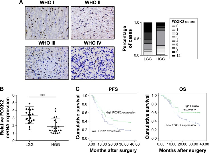
Expression of FOXK2 in different WHO grade gliomas. (A) Immunohistochemistry staining of FOXK2 in different WHO grade gliomas (original magnification ×200). The histogram shows that the expression score of FOXK2 in high-grade glioma (WHO grade III and IV gliomas) was significantly lower compared to the low-grade glioma (WHO grade I and II gliomas). The staining intensity was scored as follows: 0 (no staining), 1 (weak staining), 2 (moderate staining), and 3 (strong staining) (see Figure S1). The percentage of FOXK2-positive cells was scored as follows: 0 (0%), 1 (1%–25%), 2 (26%–50%), 3 (51%–75%), and 4 (76%–100%). (B) A total of 23 low-grade glioma tissues and 23 high-grade glioma tissues were subjected to quantitative reverse transcription PCR. The results showed that the FOXK2 mRNA level in the high-grade glioma tissues was significantly lower than in the low-grade glioma tissues. ***p<0.001. (C) Kaplan–Meier analysis for the PFS and OS based on the FOXK2 expression in the glioma patients (log-rank test, p<0.01). Blue indicates low FOXK2 expression group; green indicates high FOXK2 expression group.
Abbreviations: FOXK2, Forkhead box K2; HGG, high-grade glioma; LGG, low-grade glioma; OS, overall survival; PCR, polymerase chain reaction; PFS, progression-free survival; WHO, World Health Organization.

