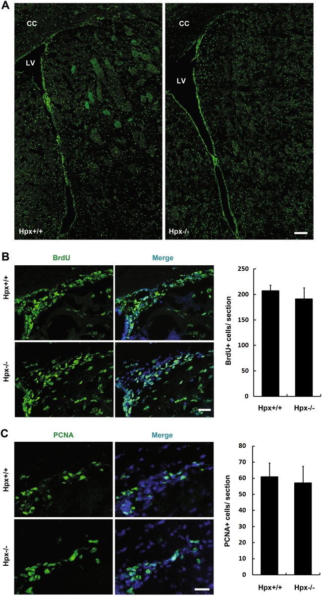
Fig. 2. Proliferation of stem cells/progenitors in SVZa was not changed by hemopexin deletion.
a Mice were dosed with BrdU and killed 2 h later. Every fifth coronal section from each brain was labeled with anti-BrdU (green) to analyze proliferation. b High-magnification images showing the BrdU-positive cells in the SVZa. Hoechst staining (blue) was used to identify nuclei. Quantification of BrdU+ cells in the SVZa (right). No significant difference was found between Hpx+/+ and Hpx−/− mice. n = 4. c High-magnification images showing the PCNA-positive cells in the SVZa. Hoechst staining (blue) was used to identify nuclei. Quantification of PCNA+ cells in the SVZa (right). No significant difference was found between Hpx+/+ and Hpx−/− mice. n = 3. Scale bar: 100 μm in a; 20 μm in b, c. LV lateral ventricle, CC corpus callosum

