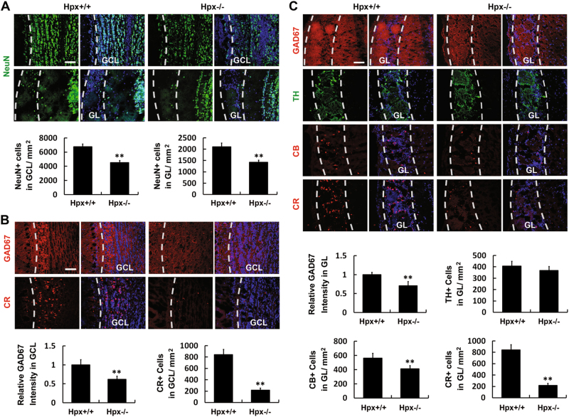
Fig. 8. Hemopexin deletion impairs the genesis of interneurons in the OB.
Coronal sections of the OB of Hpx+/+ and Hpx−/− mice were analyzed. a Anti-NeuN immunostaining (green) was performed to label the neurons. Hoechst staining (blue) was used to identify all nuclei and overall structure. Hemopexin deletion reduced the density of NeuN+ neurons in both the GCL and the GL of the OB. b Anti-GAD67 (red) and anti-CR (red) were used to label GABAergic neurons and CR+ interneurons, respectively, in the GCL of the OB. Immunoreactivity for GAD67 and the cell density of CR+ cells in the GCL were significantly reduced in Hpx−/− mice. c Anti-GAD67 (red), anti-TH (green), anti-CB (red), and anti-CR (red) were used to label GABAergic neurons, dopaminergic neurons, CB+ interneurons and CR+ interneurons, respectively, in the GL of the OB. Immunoreactivity for GAD67 and the cell density of calbindin+ (CB+) and calretinin (CR+) interneurons were decreased in the GL in Hpx−/− mice, while the cell density of TH+ dopaminergic neurons in the GL was not affected. n = 5. **p < 0.01. Scale bar: 100 μm. GCL granule cell layer, GL glomerular layer

