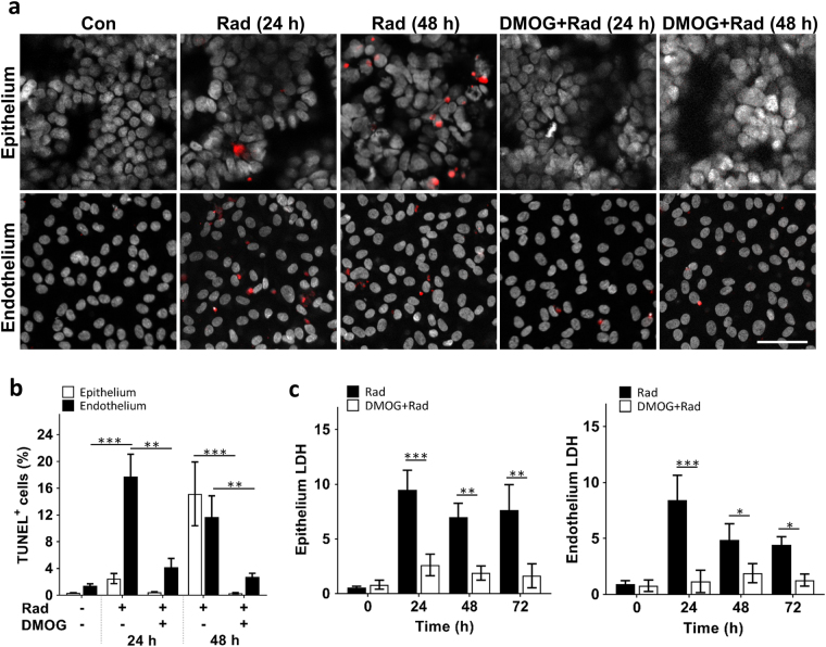
Fig. 2. Radiation-induced apoptosis and cytotoxicity in intestinal epithelium and vascular endothelium, and radio-protective effects of DMOG.
a Representative immunofluorescence micrographs of TUNEL (red) and DAPI (white) staining in epithelial and endothelial cells cultured on-chip in the absence (Con) or presence (Rad) of 8 Gy of γ-radiation (Rad), with or without DMOG treatment, 24 h and 48 h after radiation exposure (bar, 50 μm). b Graph showing the quantification of the percentage of epithelium and endothelium cells that expressed TUNEL staining (TUNEL+ cells) 24 h and 48 h after exposure to the conditions shown in a (n = 3; *P < 0.05, **P < 0.01, ***P < 0.001). c Graph showing radiation-induced cell death in the epithelium (left) and endothelium (right) in the absence (Rad) or presence of DMOG (DMOG + Rad), as assessed by quantifying LDH release from cells (data are presented as fold change in LDH levels relative to the non-irradiated control cells; n = 3; *P < 0.05, **P < 0.01, ***P < 0.001)

