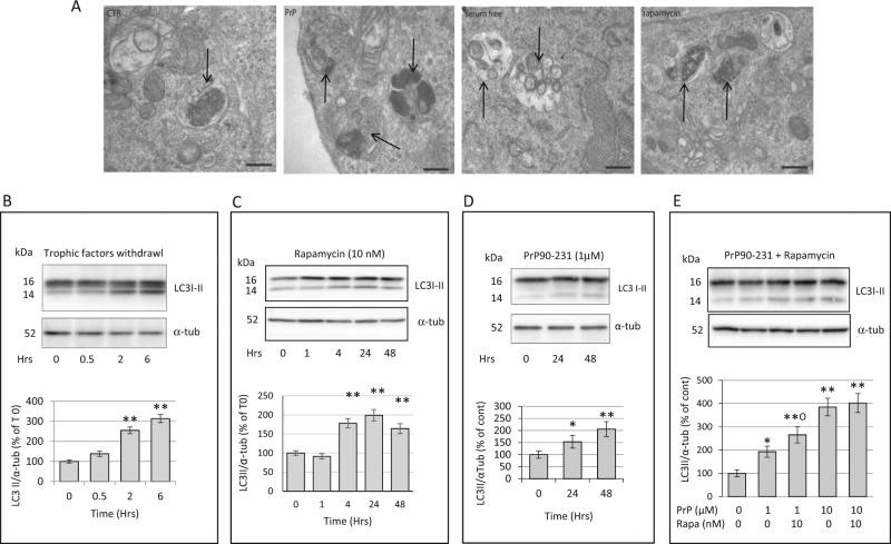
Fig. 4. Trophic factor deprivation, rapamycin and PrP90-231 increase the number of autophagosomes.
a Morphological analysis of A1 cytoplasm by electron microscopy after trophic factors withdrawal (serum deprivation), treatment with rapamycin 10 nm and PrP90-231 1 µM for 24 h. Images evidenced that serum deprivation and the treatment with both rapamycin and PrP90-231 induced appreciable increase of double-membrane autophagosomes, along with autophagolysosomes, containing electron-dense material (arrows). Space bar: 200 nm. Evaluation by immunoblotting of the time-dependent expression of LC3I-II after trophic factors withdrawal (b), rapamycin (10 nM) (c), PrP90-231 (1 μM) (d), and PrP90-231 (1 and 10 μM) in the absence or the presence of rapamycin (10 nM) (e). The amount of autophagosome-bound 14 kDa form of LC3B (LC3BII) was quantified by densitometry (histograms below each blot) and expressed as ratios on α-tubulin expression. LC3BII/α-tubulin values were expressed as percent of time 0 from three separate experiments. PrP90-231, trophic factor withdrawal, and rapamycin induced significant increase in LC3BII. *P < 0.05 and **P < 0.01 vs. time 0. Additive effects of rapamycin on increasing LC3II formation are appreciable only on PrP90-231 (1 μM). **P < 0.01 vs. cont; °P < 0.05 vs. PrP90-231. All the data are reported as mean+/− SEM of three independent experiments each performed in quadruplicate

