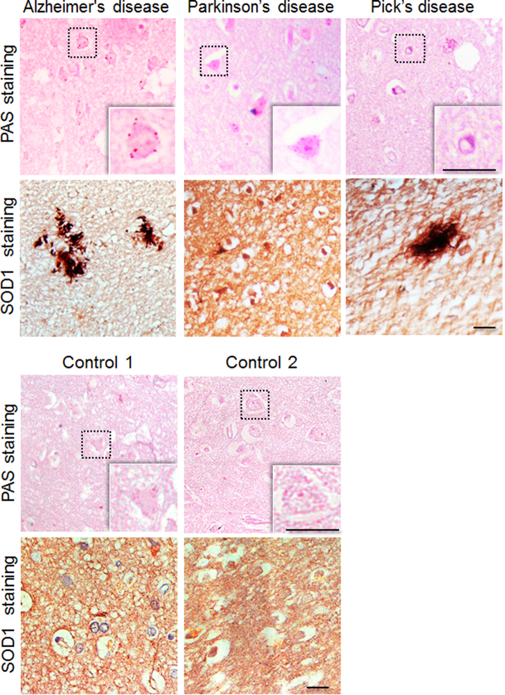
Fig. 8. Glycogen inclusions in the frontal cortex region of subjects with neurodegenerative disorders.
Representative bright field images showing PAS-positive glycogen granules in the frontal cortex region of autopsied brain tissues of subjects clinically diagnosed to have Alzheimer’s disease, Pick’s disease, or Parkinson’s disease as identified. The same regions of the brain were also immunostained with the SOD1 antibody, revealing increased levels of immunoreactivity. The panel below shows PAS and SOD1 immunoreactivity of the frontal cortex area of the two age-matched controls. These sections were from autopsied brain tissues of subjects who did not suffer from neurodegenerative conditions. Details on the subjects are given in the Supplementary Table S1. The PAS-positive glycogen granules shown here did not show autofluorescence, indicating they are not lipofuscin granules (scale bar, 10 µm)

