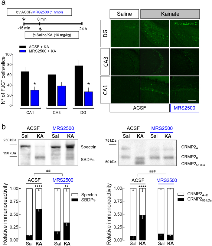
Fig. 2. Pharmacological blockade of P2Y1Rs attenuates seizure-induced neurodegeneration in the rat hippocampus.
a The intraperitoneal (i.p.) administration of 10 mg/kg of kainate (KA) in rats caused convulsive period of circa 2 h, which induced hippocampal neuronal death 24 h later, measured by FluoroJadeC-positive cells (FJC+), both in CA1, CA3, and dentate gyrus (DG) regions, as depicted in the representative images (scale bar, 200 µm) and quantified in the histogram. The hippocampi of animals injected with saline were absent of FJC+ cells. The number of FJC+ cells induced by KA was significantly attenuated by the prior i.c.v. injection of 1 nmol MRS2500 (15 min before KA injection), in DG and CA1, also showing a reduction tendency in CA3 region (p = 0.0815). The data are expressed as the number of FJC+ cells per slice (mean ± SEM) quantified from 4 animals per group and analyzing 12 coronal sections separated successively by 240 µm representing the entire hippocampus from each animal. *p < 0.05 one-way ANOVA with Sidaks’s test. b Western blot of calpain substrates spectrin and CRMP2 in hippocampal protein extracts prepared 24 h after saline/KA injection. KA-injected animals displayed an increase of the percentage of spectrin breakdown products (SBDPs) with an apparent molecular weight of ~145 kDa, and of the truncated form of CRMP2 (CRMP255 kDa). The prior injection of MRS2500 attenuated or prevented the calpain-mediated cleavage of spectrin or CRMP2, respectively. Data are mean ± SEM of the relative percentage of Spectrinfull-length/SBDPs and CRMP2A+B/CRMP255 kDa (n = 4). **p < 0.01 and ****p < 0.0001 one-way ANOVA with Sidak’s test for KA vs. saline. ##p < 0.01 and ###p < 0.001 two-way ANOVA

