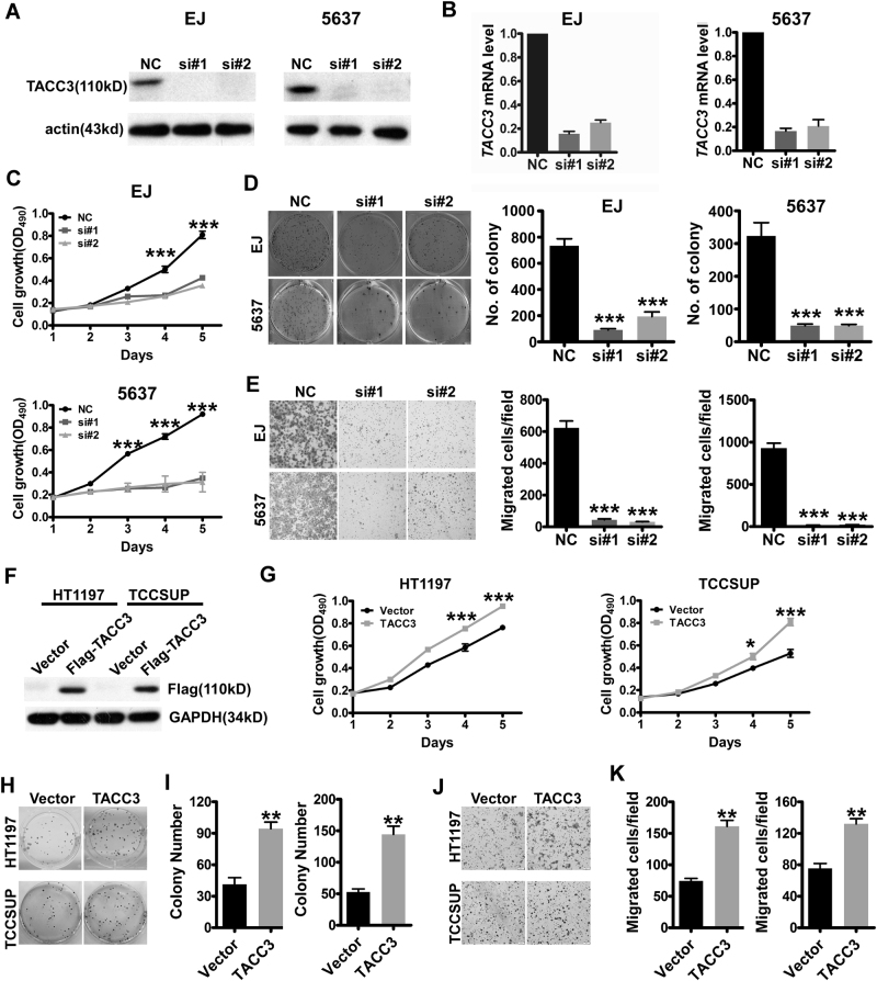
Fig. 2. Alternative expression of TACC3 regulates cell growth and invasion in bladder cancer.
a, b Western blotting (a) and QPCR (b) analyses show almost complete elimination of TACC3 expression after TACC3 siRNA knockdown in EJ and 5637 cells. c Suppression of TACC3 in EJ and 5637 cells dramatically reduced their proliferative ability, as determined by MTT assay. ***p < 0.001 d TACC3 knockdown in EJ and 5637 cells markedly reduced their ability to form foci, as determined by the foci-formation assay. Left: representative images of colony formation. Right: error bars present means ± SEM (n = 3). p-Values were calculated by one-way ANOVA. **p < 0.01. e Transwell assay showed downregulated TACC3 reduced cell invasiveness. Left: representative images. Right: error bars present means ± SEM (n = 3). p-Values were calculated by one-way ANOVA. ***p < 0.001. f Levels of ectopically expressed TACC3 in HT1197 and TCCSUP cells were examined by western blotting. g Overexpression of TACC3 promoted cell growth, as shown by MTT assay. ***p < 0.001. h, i Overexpression of TACC3 increased the formation of foci, as determined by the foci-formation assay. j, k Overexpression of TACC3 enhanced cell invasiveness. The statistical results in i and k are reported as mean ± SEM. p-Values were calculated by the Student’s t-test. **p < 0.01

