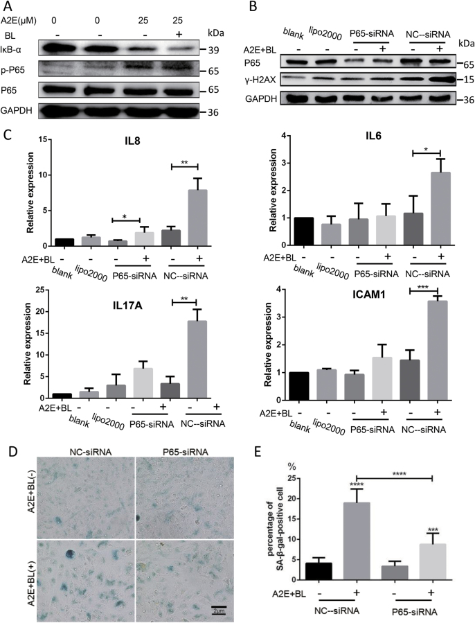
Fig. 5. Photosensitization of A2E triggered SASP through NF-κB pathway activation.
a Western blot assay targeting IκB-α, p-p65, and p65 proteins in RPE cells treated with 25 μM A2E under photosensitization. b Western blot assay targeting p65 and γ-H2AX in RPE cells with suppression of p65 functions. c Expression of IL8, IL6, IL17A, and ICAM1 in RPE cells with suppression of p65 functions determined by RT-qPCR. Expression is normalized to the expression of β-actin and is expressed as the fold change relative to the control. Values are presented as the means ± SD of two independent samples with triplicate qPCR; * indicates p value < 0.05, ** indicates p value < 0.01, *** indicates p < 0.001. d Representative microscopic images of β-galactosidase staining in RPE cells seeded in cell culture carrier plates. RPE cells seeded in cell culture inserts treated with 25 μM A2E under photosensitization with suppression of p65 functions. PDL = 15. e Quantification of the percentage of cells with positive SA-β-gal staining shown in d; **** indicates p < 0.0001 compared to control. The experiment was performed independently at least three times

