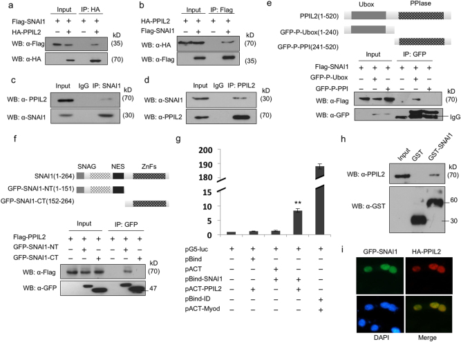
Fig. 3. PPIL2 interacted with SNAI1.
a, b Co-IP assay showed the interaction between exogenous PPIL2 and SNAI1 in T47D cells. c,d Co-IP assay showed the interaction between endogenous PPIL2 and SNAI1 in T47D cells. e Co-IP assay showed that PPIL2 interacted with SNAI1 via its U-box domain in T47D cells. f Co-IP assay showed that SNAI1 interacted with PPIL2 via its N terminal in T47D cells. g The direct interaction between PPIL2 and SNAI1 was observed using mammalian two-hybrid system. h GST pull-down assay showed the interaction between GST-SNAI1 and endogenous PPIL2 in MCF cells. i Immunofluorescence showed that PPIL2 (red) and SNAI1 (green) colocalized in the nucleus of MCF-7 cells (20×)

