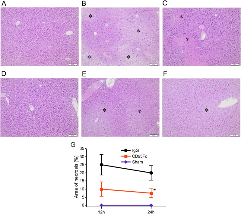
Fig. 3. Hepatocellular necrosis is ameliorated by the application of CD95Fc.
Liver injury was induced by 45 min of warm ischemia of the median and left lateral liver lobe after prior application of two IV injections of CD95Fc (30 mg/kg) or the same volume of IgG (30 mg/kg) antibody at 12 h and at 30 min before ischemia. After 12 h and again after 24 h of reperfusion, liver tissue was harvested and processed for histopathology. Representative H&E-stained liver sections (×100 magnification, areas of necrosis marked with an asterisk). a sham, reperfusion 12 h; b IgG, reperfusion 12 h; c CD95Fc, reperfusion 12 h; d sham, reperfusion 24 h; e IgG, reperfusion 24 h; f CD95Fc, reperfusion 24 h. g Area of necrosis (% of area) (mean ± S.D., n = 6. *p < 0.0001, MANOVA, between-subject-effect compared to IgG, time points 12 and 24 h were considered as repeated measurements)

