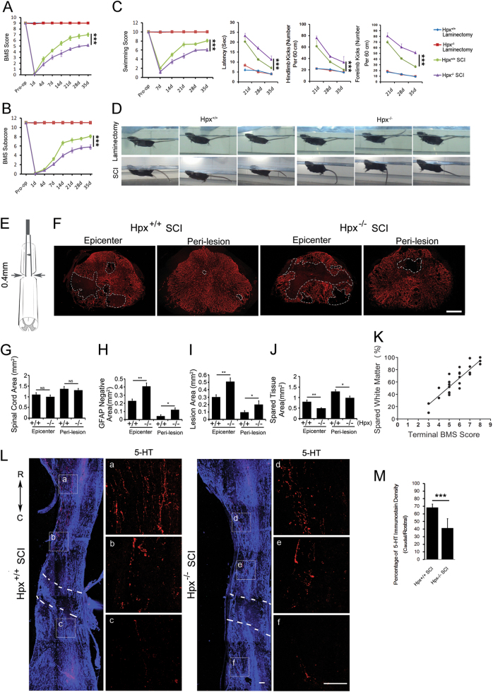
Fig. 4. Hemopexin knockout mice showed reduced functional recovery of hindlimb after SCI.
Functional recovery was assessed by determining the Basso Mouse Scale (BMS) scores (a), Basso Mouse Scale subscores (b) and swimming tests (c, d) over a 35-day period (n = 15 mice per group). e Schematic graph of the thoracic spinal cord (T8–T9) crush model used. f Representative images of GFAP immunostaining in spinal cord sections at the epicenter and peri-lesion sites from Hpx−/− and Hpx+/+ mice on 35 dpl after SCI. The white line denotes the size of the lesion. Quantification of GFAP immunostaining in the total tissue area (g), GFAP-negative area (h), lesion area (i), and spared tissue (j). The relationship between spared white matter and final BMS score of each mouse was assessed with Pearson’s regression analysis (k). l Representative images of 5-HT+ fibers (red) co-stained with GFAP (blue) in sagittal sections. m Quantification reveals a significant decrease in 5-HT+ fiber sprouting caudal to the injury in Hpx−/− SCI group vs. Hpx+/+ SCI group mice on 35 dpl. a–f 5-HT+ fibers in the boxed areas are enlarged in the right panels in two groups. Dashed lines indicate lesion margins. (n = 5 mice per group). *p < 0.05, **p < 0.01, ***p < 0.005, compared with the indicated control. Scale bar = 100 µm

