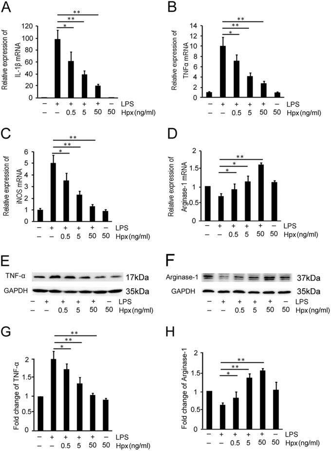
Fig. 5. Hpx switches M1 microglia to the M2 polarization state in vitro.
a–d Effects of Hpx on the polarization of LPS-stimulated microglia (MGLPS) were determined using quantitative RT-PCR. IL-1β, iNOS, and TNF-α were used as M1 markers (a–c) and Arg-1 was used as an M2 marker (d). e–h Western blot analysis of TNF-α (e) and Arg-1 (f) expression in LPS-stimulated microglia. g, h Scanning densitometry of TNF–α (g) and Arg-1 (h) levels, which were quantified and normalized to GAPDH. *p < 0.05, **p < 0.01 compared with the indicated control. Data are presented as the means ± SEM of at least three independent experiments

