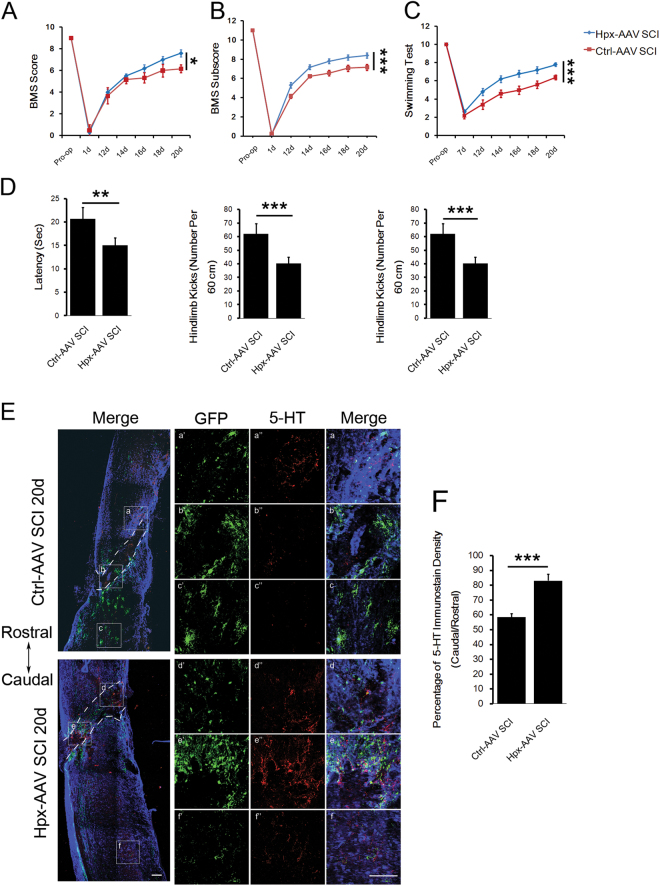
Fig. 8. Hpx promoted functional recovery and raphespinal sprouting in lesion spinal cord of the Hpx-AAV SCI group.
Functional recovery was assessed by determining BMS scores (a), BMS subscores (b), and swimming tests (c, d) between 2 and 3 weeks postsugery (N = 10 mice per group). e Representative images of 5-HT+ serotonergic fibers (red) in the middle panels, costained with GFAP (blue) and GFP (green) in sagittal sections. Green indicates expression of GFP fused with or without Hpx expressed by AAV vectors injected into the spinal cord at the lesion. a–f Boxed areas are enlarged in the right panels (a’–f’, a”–f”). f Quantification reveals a significant increase in 5-HT+ fiber sprouting rostral to the injury in the Hpx-AAV SCI group vs. Ctrl-AAV SCI group of mice on 20 dpl. Dashed lines indicate lesion margins. Scale bar = 100 μm; *p < 0.05, **p < 0.01, ***p < 0.005

