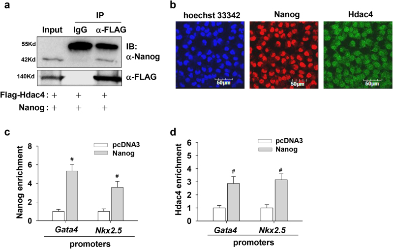
Fig. 7. Functional association between Nanog and Hdac4 inhibits Gata4 and Nkx2.5 expression.
a Immunoprecipitation assay shows the functional interaction of Nanog and Hdac4. 293T cells were transfected with expression vectors for Nanog and FLAG-Hdac4. Cell lysates were subjected to immunoprecipitation with anti-FLAG antibody or normal IgG as a negative control, and the precipitates were subjected to immunoblot analysis with anti-Nanog or anti-FLAG antibodies, respectively. Five percent of the whole-cell lysate was loaded for the input control. b Immunostaining of undifferentiated P19 cells with specific antibodies shows the subcellular localization of Nanog and Hdac4. Hoechst dye was used for staining nuclei. c, d Nanog overexpression facilitates the recruitment of Hdac4 onto Gata4 and Nkx2.5 promoters. Undifferentiated P19 cells were transfected with plasmids encoding Nanog. After 48 h of transfection, cells were collected and ChIP assays were conducted with antibodies against Nanog (c) and Hdac4 (d). Immunoprecipitated DNA fragments were amplified by real-time PCR for the promoter regions of Gata4 and Nkx2.5 (S1 region). Each bar represents mean ± SD from three independent experiments (*p < 0.05; #p < 0.01 vs. the control)

