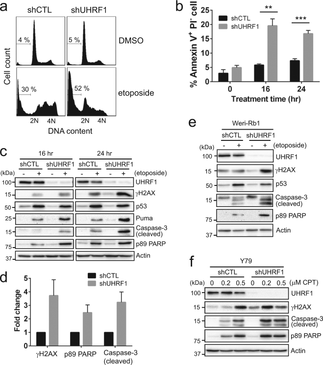
Fig. 2. Enhanced drug sensitivity upon UHRF1 depletion involves apoptotic cell death.
a Sub-G1 population detected by flow cytometry in control (shCTL) and UHRF1-knockdown (shUHRF1) Y79 cells treated with vehicle or 10 µM etoposide for 24 h. The percentage of sub-G1 population is shown. b Quantification of early apoptotic cell death by Annexin V-PI staining. Control and UHRF1-knockdown Y79 cells were treated with 10 µM etoposide for the time indicated. The data represent the mean ± SD of % Annexin V+ PI− population from triplicate experiments. **P < 0.01, ***P < 0.001. c Immunoblots for indicated proteins in Y79 shCTL and shUHRF1 cells after exposure to 10 µM etoposide for 16 h and 24 h. d Densitometric analyses of the indicated proteins for 24 h-treatment in c. The graph is shown as the mean ± SD of fold changes from four independent experiments, relative to the normalized level in shCTL cells. e Immunoblots for indicated proteins in Weri-Rb1 shCTL and shUHRF1 cells treated for 24 h. f Expression of indicated proteins in UHRF1-knockdown Y79 cells after treatment with various concentrations of camptothecin (CPT) for 24 h

