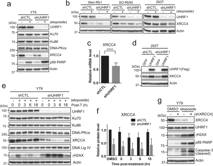
Fig. 4. XRCC4 downregulation in UHRF1-depleted retinoblastoma cells impedes recovery from DNA damage.
a Immunoblots for indicated proteins in shCTL and shUHRF1 Y79 cells. Cells were treated with 10 µM etoposide for 24 h. b Expression of XRCC4 in UHRF1-depleted Weri-Rb1, SO-Rb50, and 293T cells after treatment with 10 µM etoposide for 24 h. c qRT-PCR analysis of relative XRCC4 expression in Y79 shCTL and shUHRF1 cells. The bar graph is shown as the mean ± SD of fold changes from five independent experiments, relative to the normalized XRCC4 expression in control-knockdown cell. ****P < 0.0001. d XRCC4 expression after adenoviral expression of either GFP or Flag-tagged UHRF1 in shCTL and shUHRF1 293 T cells. e Immunoblots for indicated proteins showing the recovery kinetics from acute DNA damage induced by etoposide. Cells were treated with 10 µM etoposide for 1 h, and then placed in fresh media without drugs for the indicated time post-treatment (post-T). f Densitometric analysis of XRCC4 protein levels in e. The graph is shown as the mean ± SD of fold changes from five independent experiments, relative to the normalized level in DMSO-treated shCTL Y79 cells. g Immunoblots for indicated proteins in shCTL (−) and shXRCC4 (+, clone #40116) Y79 cells after treatment with vehicle or 10 µM etoposide for 24 h

