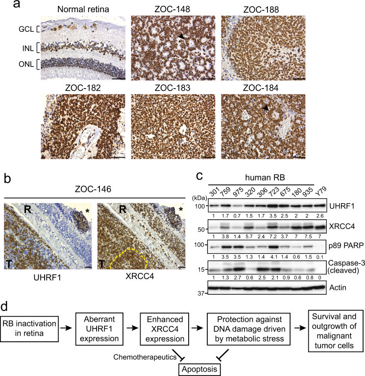
Fig. 6. XRCC4 expression in human primary retinoblastoma.
a Immunostaining of XRCC4 in human retinoblastoma (n = 10) and normal retina (42 years of age) sections. Nuclei were counterstained with hematoxylin. Black arrowheads in ZOC-148 and ZOC-184 indicate rosettes characteristic of differentiated retinoblastoma. GCL ganglion cell layer, INL inner nuclear layer, ONL outer nuclear layer, Scale bar: 50 µm. b Expression of UHRF1 and XRCC4 in human retinoblastoma. Two serial sections of ZOC-146 immunostained for UHRF1 and XRCC4 are shown as representative images. Dense staining of XRCC4 is visible in tumor foci in the retina (marked by *) and vitreous tumor region (marked by yellow dashed lines) where UHRF1 is highly expressed. T tumor, R retina, scale bar: 50 µm. c Immunoblots for indicated proteins in human retinoblastoma (RB) lysates. Relative abundance of proteins determined by densitometry is shown below each panel. d Proposed model of UHRF1-mediated tumor promotion in retinoblastoma

