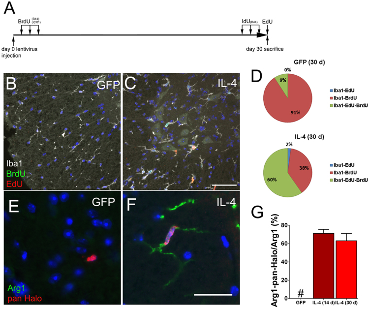
Fig. 3. IL-4 increases S-phase tracers’ incorporation in Arg1 expressing cells.
a Schematic representation of the labeling paradigm. b and c Representative triple immunofluorescence for Iba1, BrdU and EdU from GFP-LV and IL-4-LV mice collected at day 30. d Quantifications of Iba1/ BrdU/ EdU positive cells on GFP-LV mice (n = 4) and IL-4-LV mice (n = 6), (GFP: Iba-EdU 0%; Iba-BrdU 91 ± 5%; and Iba-EdU-BrdU 9 ± 5%); (IL-4: Iba-EdU 2 ± 1.6%; Iba1-BrdU 38 ± 25%; and Iba1-EdU-BrdU 60 ± 26%). e and f Double labeled sections for Arg1 and an anti- pan Halogen antibody from GFP-LV and IL-4-LV mice. g Percentages (mean ± S.D.) of double positive Iba1/Arg1 cells in IL-4-LV mice (14d n = 5, 30d n = 3). GFP-LV mice (n = 5) did not show any double positive cells. One-way ANOVA followed by Bonferroni’s multiple Comparison test has been used to analyze data in (g). Scale bar 50 µm in (c) and 10 µm in (f). # not detectable

