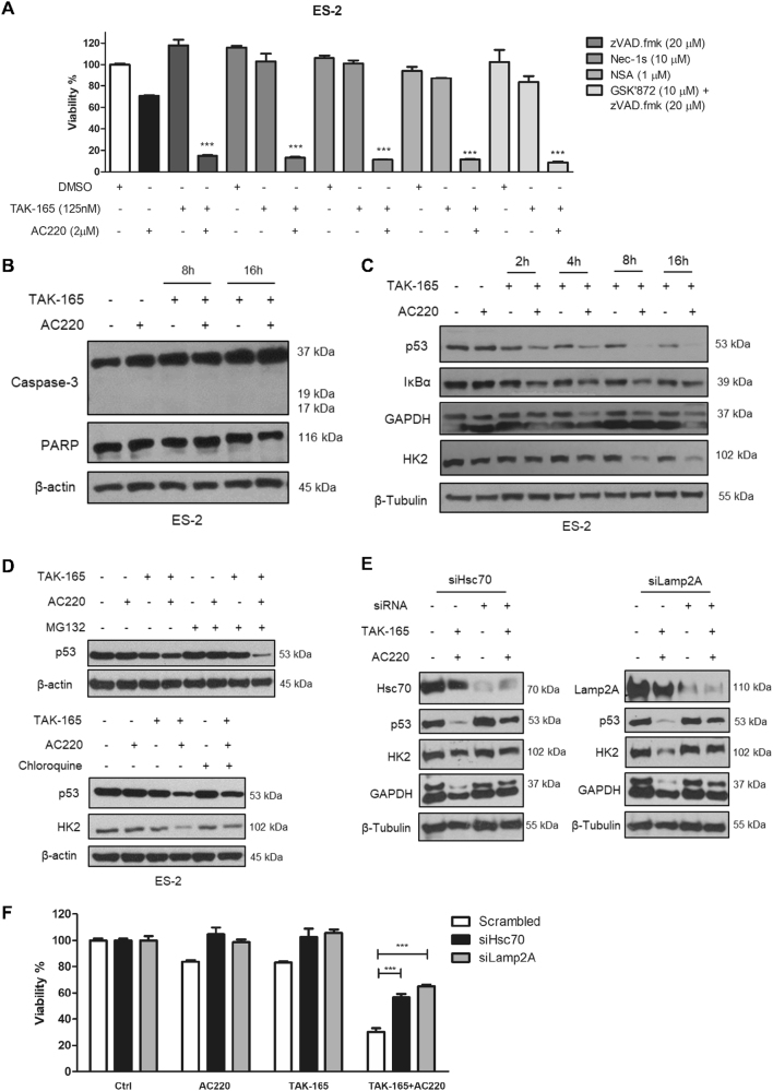
Fig. 6. TAK-165/AC220 induces cell death through Chaperone-mediated autophagy activation.
a Cell viability (%) of ES-2 cells treated with treated with TAK-165, AC220 or a combination of TAK-165 plus AC220 in the presence or absence of zVAD.fmk, Nec-1s, NSA and GSK832 for 24 h (n = 3). Viability was determined using CellTiter-Glo® Luminescent assay. b Immunoblotting of caspase-3 and PARP-1 in ES-2 cells treated with TAK-165 (125 nM) and/or AC220 (2 μM) for 8 and 16 h. c TAK-165/AC220 combination activates chaperone-mediated autophagy. Immunoblotting of p53, IκB-α, GAPDH and HK2 levels in ES-2 cells treated with TAK-165 and/or AC220 up to 16 h. d Immunoblotting of p53 levels in ES-2 cells treated with TAK-165 and AC220 for 6 h in the absence or presence of MG132 (10 μM, proteasome inhibitor) and Chloroquine (CQ, 50 μM). e Immunoblotting of Lamp2A, Hsc70, p53, HK2 and GAPDH levels in Scramble, Hsc70, or Lamp2A siRNA-transfected ES-2 cells treated with TAK-165 (125 nM) and AC220 (2 μM) for 8 h. f Cell viability (%) of scramble, Lamp2A, or Hsc70 siRNA-transfected ES-2 cells treated with TAK-165 and/or AC220 for 8 h. Viability was determined using CellTiter-Glo® Luminescent assay (n = 3). Anti–β-tubulin and anti-β-actin were used as a loading control. In all the experiments, treatment groups were compared with control group, unless otherwise indicated. Bars: Mean ± SD. ***p < 0.001

