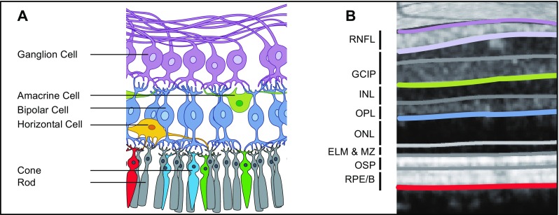
Fig. 1.

Anatomy of the retina (a) with corresponding layers measured by OCT as suggested by Staurenghi et al. [172] and Cruz-Herranz et al. [97] (b). Parts of the figure are provided by courtesy of www.neurodial.de [173]. OCT optical coherence tomography, RNFL retinal nerve fiber layer, GCIP combined ganglion cell and inner plexiform layer, INL inner nuclear layer, OPL outer plexiform layer, ONL outer nuclear layer, ELM & MZ external limiting membrane and myoid zone, OSP outer segments of photoreceptors (ellipsoid zone), RPE/B retinal pigment epithelium and Bruch’s complex
