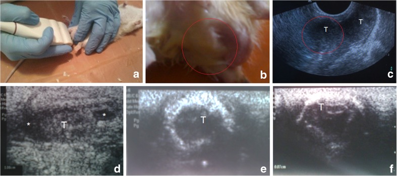
Fig. 3.
Assessment of the rat testes: a – view of ultrasound technique to obtain transversal scans of testes in rat; b, c – acquired cryptorchidism under LF model – general view (b) and corresponding US image (c), whereas red circles indicate the right testicle (T) not descended to the scrotum; d, e, f – longitudinal US scan of testes: decreasing size, fibrosis, deformation, petrification, stiff and thick (fibrotic) capsule, irregular contour; * – fluid around a testicle (hydrocele)

