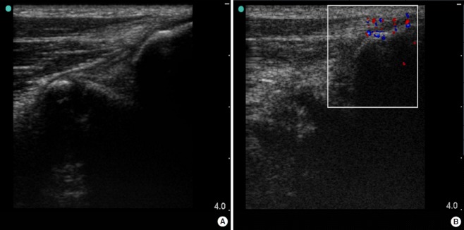
Fig. 1.
Result of sonography. Medial elbow tendinopathy and insertion of common flexor origin at medial epicondyle, in a longitudinal view: (A) Grayscale B-mode ultrasound shows irregularity and a focal hypoechoic area in the common flexor origin, (B) Color Doppler ultrasound shows increased blood flow inside the hypoechoic area. The red color dots show the blood flow towards the transducer, and blue ones show the blood flow away from the transducer.

