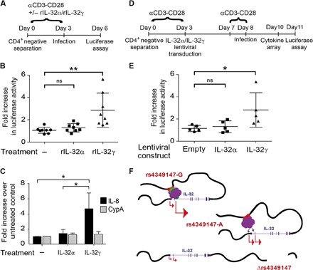
Fig. 4. Relative ratio of IL-32 isoforms influences susceptibility of primary CD4+ T cells to HIV infection and alters the proinflammatory cytokine environment.

(A) CD4+ T cells isolated from eight healthy donors were stimulated with CD3-CD28–coated beads in the presence or absence of rIL-32α or rIL-32γ and infected with an HIV virus harboring a luciferase reporter, as shown in the schematic. Efficiency of infection was determined by measuring luciferase activity 72 hours after infection. (B) CD4+ T cells exogenously incubated with rIL-32γ are more susceptible to HIV infection than mock-treated cells and cells treated with rIL-32α. ns, not significant. (C) Real-time PCR analysis demonstrates that CD4+ T cells incubated with rIL-32γ, but not those treated with rIL-32α, show increased IL-8 expression. CypA, cyclophilin A. (D) CD4+ T cells isolated from five healthy donors (rs4349147-G/A) were stimulated with CD3-CD28–coated beads and infected with a control lentiviral vector or a lentiviral vector expressing IL-32α or IL-32γ. After 4 days, the cells were restimulated for 24 hours and infected with an HIV virus harboring a luciferase reporter, as shown in the schematic. Efficiency of infection was determined by measuring luciferase activity 72 hours after infection. (E) Exogenous lentiviral overexpression of IL-32γ results in an increase in HIV infection as compared to CD4+ T cells overexpressing IL-32α or control cells infected with an empty lentiviral construct. (F) Model depicting rs4349147 regulation of IL-32 expression. See Discussion for explanation.
