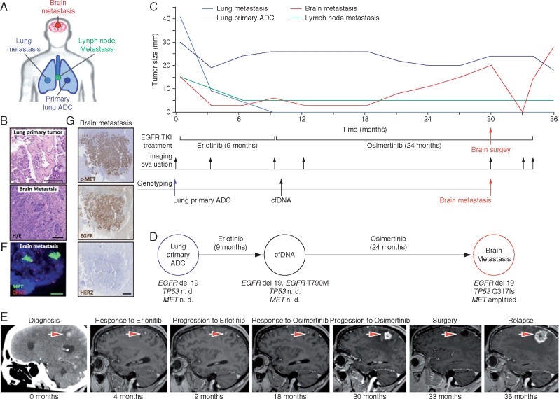
Figure 1.
Evolution and plasticity of acquired resistance mechanisms to osimertinib in NSCLC harboring EGFR mutation. (A) Study of the molecular profiling of metastatic brain biopsy specimen of female patient with NSCLC exon 19 deletion and T790M mutation treated with osimertinib. (A, C and D) ADC, adenocarcinoma. (B) Morphological appearance of primary and metastatic lung lesions (haematoxylin and eosin, 20×). (C) Serial of target tumor lesions measures and the lower panel displays anti-EGFR treatment, imaging evaluation and genotyping along the evolution of the metastatic disease. (D) Molecular profiling of paired biopsies: baseline and at the time of progression to erlotinib and osimertinib. n. d., non-determined. (E) Representative brain MRI and CT scans at the time points indicated are provided; the largest brain target lesion is indicated with an arrow. (F) FISH analyses showing the presence of MET amplification in the brain metastasis after relapse osimertinib (MET gene, green signals; CEN7, red signals; 100×). (G) High expression of cMET and EGFR proteins was observed in brain lesion by immunohistochemistry. No expression for HER2 was found (2.5×).

