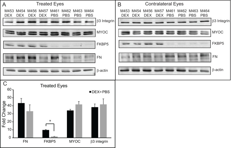
Fig 4. Western blotting of lysates from anterior segments of mouse eyes treated with DEX or PBS for 5 weeks.
(A) Western blots of lysates from eyes treated with DEX or PBS for 5 weeks. (B) Western blot of lysates from the contralateral untreated eyes of the same mice as in (A). (C) Densitometry of western blots shown in (A), normalized to the β-actin loading control. DEX treated versus PBS treated eyes were significantly different, *p<0.05.

