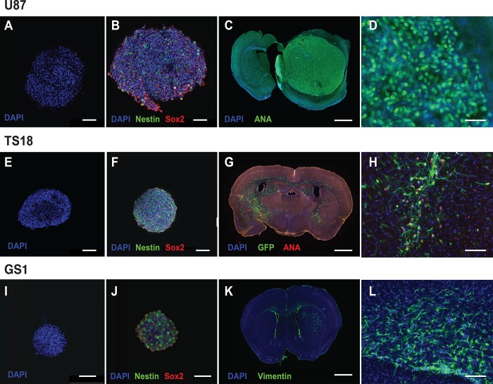
Fig 1. Cell line characterization demonstrated stem-cell characteristics and tumorigenicity in vivo.
(A-B) U87 neurosphere culture demonstrating Nestin (green) and Sox2 (red) staining in serum-free culture (s.b. = 100μm). (C) U87 orthotopic implantation model showing large well-defined masses within the brain parenchyma, showing staining for anti-human nuclear antigen (green) (s.b. = 1000μm). (D) High power field (x40) of well-demarcated tumour edge showing anti-human nuclear antigen staining of tumour cells (s.b. = 50μm). (E-F) TS18 in serum free neurosphere culture showing uniform Nestin and Sox2 staining (s.b. = 100μm). (G) Orthotopic implantation of TS18-GFP cells with combined GFP (green) and anti-human nuclear antigen (red) staining to show diffuse infiltration of cells both within the parenchyma and across the corpus callosum into the opposite hemisphere (s.b. = 1000μm). (H) High power field (x20) showing area of diffuse infiltration and co-expression of GFP and anti-human nuclear antigen in implanted cells (s.b. = 100μm). (I-J) GS1 neurosphere culture showing uniform Nestin and Sox2 staining in serum free culture. (K) Orthotopic implantation of GS1 cells into the striatum with anti-vimentin staining. Vimentin staining is characteristic of gliosarcoma and is not found in the parenchyma, and was used to trace GS1 cells within the parenchyma (s.b. = 1000μm). (L) High power field (x20) showing diffusely infiltrating vimentin positive cells (green) within the corpus callosum (s.b. = 100μm).

