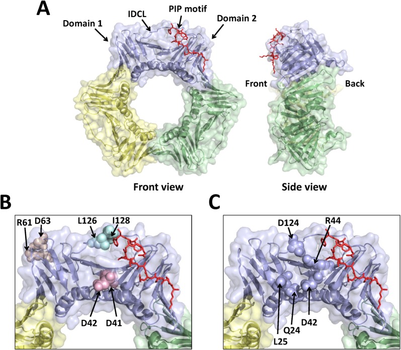
Fig 1. Location of the three double alanine substitutions in PCNA.
(A) Front and side views of the wild-type PCNA trimer (PDB entry 2OD8.pdb). The three subunits of PCNA are colored light blue, pale green, and pale yellow. The bound PIP motif is shown in the stick representation and colored red. (B) Close-up view of one subunit of PCNA with the location of each substituted amino acid residue shown in the sphere representation. The D41A/D42A, R61A/D63A, and L126A/I128A substitutions are colored light pink, wheat, and pale cyan, respectively. (C) Close-up view of one subunit of PCNA with the location of the residues comprising the surface cavity shown in the sphere representation.

