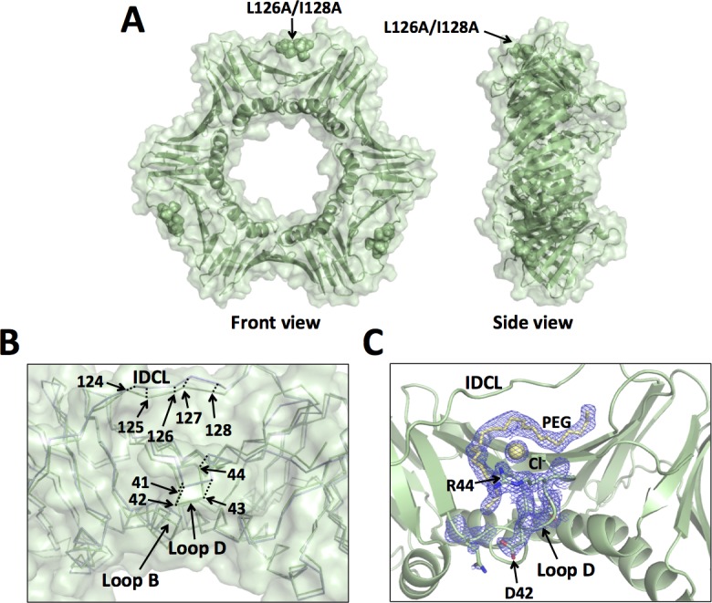
Fig 5. X-ray crystal structure of the L126/I128A mutant PCNA protein.

(A) Front and side views of the L126A/I128A mutant PCNA protein. All three subunits are colored pale green, and the locations of the substituted amino acid residues are shown in the sphere representation. (B) Close up of an overlay of the wild-type PCNA protein (light blue) and the L126A/I128A mutant PCNA protein (pale green) are shown in the ribbon representation (RMSD of 0.835 Å). The positions of the α carbons of residues 41 to 44 and of residues 124 to 128 are indicated. (C) Close up of the structure of the L126A/I128A mutant PCNA protein shown in the cartoon representation. The side chains of D42 and R44 are shown in the stick representation. A portion of a PEG molecule and a chloride ion are shown in the stick and sphere representation, respectively. The 2Fo-Fc map contoured at 1 σ is shown.
