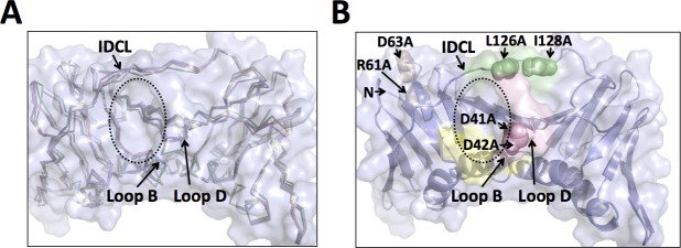
Fig 6. Comparison of the surface cavity in the wild-type and mutant PCNA proteins.
(A) Close up of an overlay of the wild-type PCNA protein (light blue), the D41A/D42A mutant PCNA protein (light pink), the R61A/D63A mutant PCNA protein (wheat), and the L126A/I128A mutant PCNA protein (pale green) are shown in the ribbon representation. The positions of loop B, loop D, and the IDCL are indicated. The edge of the surface cavity is highlighted with a dashed ellipse. (B) Close up of the structure of the wild-type PCNA protein (light blue) is shown in the cartoon representation. The locations of residues D41 and D42 (light pink), residues R61 and D63 (wheat), and residues L126 and I128 (pale green) are shown in the sphere representation.

