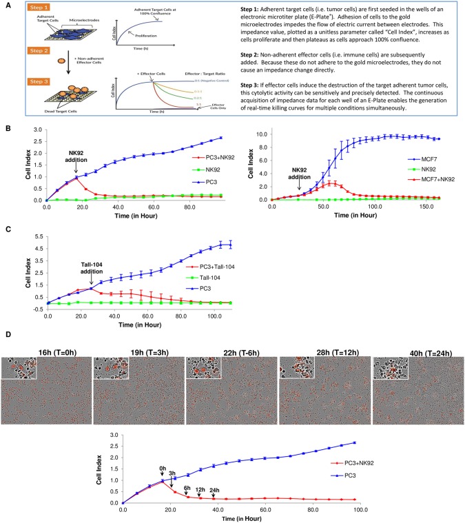
Fig 1. Working principle of the xCELLigence RTCA system and result from NK92 and TALL-104.
(A) Working principle of xCELLigence impedance technology applied to immunotherapy monitoring. The xCELLigence RTCA label-free technology monitors cell number by changes in impedance measured through gold electrodes embedded in proprietary E-Plates. When seeded alone, target adherent cancer cells proliferation rate is registered as increase in the impedance-related Cell Index (CI) parameter over time. Effector non-adherent immune cells produce small baseline level signal due absence of tight surface adhesion over the gold electrodes. When immune cells are added to adherent target cells, their cytolytic activity causes the adherent cells to round up and detach, consequently reducing CI value. (B) Impedance monitoring is validated using NK92 as effector cells over nuclear red-labeled PC3 prostate cancer cells (left) or MCF7 breast cancer cells (right) as targets. When seeded alone, target cells adhere to the plate and proliferate, increasing the CI readout (blue lines). NK92 effector cells seeded alone caused only a small increase in the CI value over the initial background measurement (green lines). When added to target cells, NK92 cause cell cytolysis and subsequent progressive decrease in CI (red lines). Y-axis is the normalized cell index generated by the RTCA software and displayed in real time. X-axis is the time of cell culture and treatment time in hour. Mean values of the CI were plotted ± standard deviation. Time interval is 6 hours for all the figures unless indicated. (C) The same PC3 target cells are treated with a cytotoxic T cell line (Tall-104), showing the suitability of the technology to use different effector cell types. (D) Images taken at different time points after NK92 addition show correlation between CI drop (red line in the plot), reduction in target cells number (red nuclei PC3 in the images) and changes in cell morphology/adhesion in apoptotic cells (red nuclei in images enlargements).

