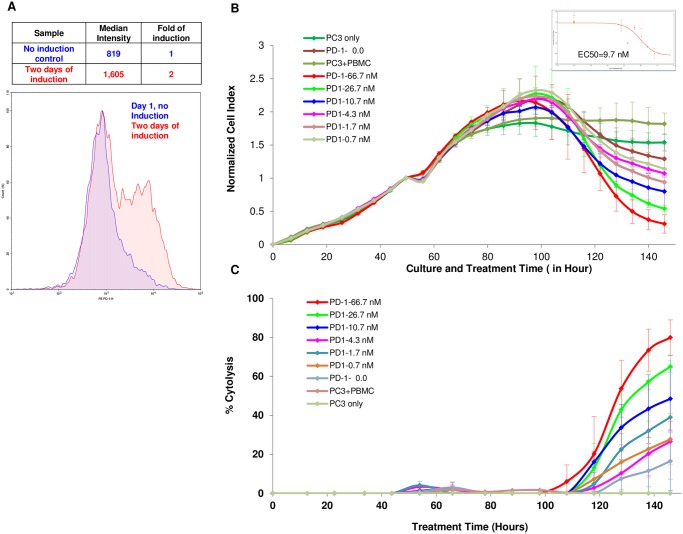
Fig 7. PC3 killing by PBMCs is enhanced by anti PD-1 blocking antibody.
(A) Freshly isolated PMBCs shows increased PD-1 expression after SEB stimulation. (B) PC3 at 5,000 cells per well were seeded on E plate and treated with freshly isolated PBMCs two days after initial seeding in presence with increasing concentrations of the anti PD-1 blocking antibody. PC3 cells only and PC3 cells plus PBMCs and without antibody were included as controls. The insert show the dose dependent curve and EC50 calculated at t = 150 hours. (C) Same data from (B) but displays as % cytolysis.

