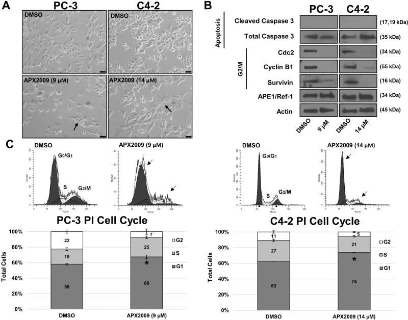
Figure 5. APE1/Ref-1 redox inhibition induces G1 cell arrest.
(A) PC-3 and C4-2 cell lines were treated with DMSO or APX2009 (9 and 14 µM, respectively) for 48 hours. Representative images were taken at 20× Magnification. Scale bar = 50 µm. (B) Immunoblotting was performed and membranes were probed with antibodies for Cleaved Caspase 3, Total Caspase, Cyclin B1, Cdc2, survivin and Actin as labeled. (C) PC-3 and C4-2 cells were treated with DMSO or APX2009 (9 and 14 µM, respectively) for 48 hrs and then collected and stained with RNAse/PI wash. Flow Cytometry was then performed. n =3, *-denoting p < 0.05 by unpaired Student’s t-test.

