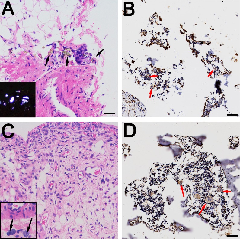
Figure 4. Histological examination of rat synovium tissues.
(A and C) Hematoxylin and eosin staining of synovium (A for PEEK, C for CoCrMo), indicated by black arrows; wear particles (A) and inflammatory cells (C) can be observed in the necrotic tissues. Polarizing microscopy of PEEK particles is shown in the lower left quarter. (B and D) Immunohistochemical staining of synovium (B for PEEK, D for CoCrMo), indicated by red arrows; multinucleated giant cells and Langhans giant cells are concentrated at the sites of wear particles. Bar = 50 μm.

