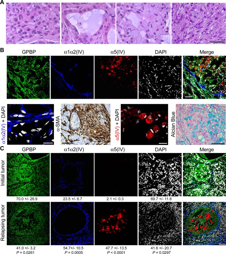
Figure 3. GPBP and mesh collagen IV networks associate with EMT phenotypes in human NSCLC.
(A) HE analysis of cell types in small A549 tumors. Left to right: poorly differentiated, epithelial, signet ring-like and fusiform cells. (B) Frozen (IF-CM) and paraffin-embedded (α-SMA and Alcian Blue) sections of small A549 tumors were analyzed. Upper, IF-CM detection of the indicated polypeptides in stromal and nodular tumor regions. Lower, from left to right the indicated polypeptides were detected by IF-CM (bar = 10 μm), horseradish peroxidase immunohistochemical method (brown) (bar = 100 μm) in tumor peri-nodular stroma, IF-CM (bar = 10 μm), and Alcian blue staining (bar = 20 μm) of the mucus (blue) in tumor nodular regions. (C) Paraffin-embedded sections of a NSCLC patient tumor at diagnosis prior to initiation of chemotherapy (Initial tumor) and at relapsing after surgery and chemotherapy (Relapsing tumor) were analyzed by IF-CM to visualize the indicated proteins. Indicated are FI measured, expressed and compared as in Figure 1. Statistics: Student’s t-test. Similar conclusions were obtained when comparing untreated vs after treatment chemoresistant NSCLC specimens.

