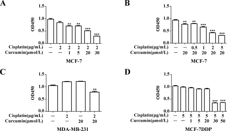
Figure 5. Curcumin’s effect on breast cancer cell sensitivity to cisplatin.
(A) MCF-7 cells were treated with 2 μg/mL cisplatin combined with increasing curcumin concentrations for 48 h, and cell proliferation was analyzed by CCK-8 assay. (B) MCF-7 cells were treated with 20 μmol/L curcumin and increasing cisplatin concentrations for 48 h, and cell proliferation was analyzed by CCK-8 assay. (C) MDA-MB-231 cells were treated with 2 μg/mL cisplatin and 20 μmol/L curcumin for 48 h, and cell proliferation was analyzed by CCK-8 assay. (D) MCF-7DDP cells were treated with 5 μg/mL cisplatin and increasing curcumin concentrations for 48 h, and cell proliferation was analyzed by CCK-8 assay. **P < 0.01, ***P < 0.001.

