
Figure 5.
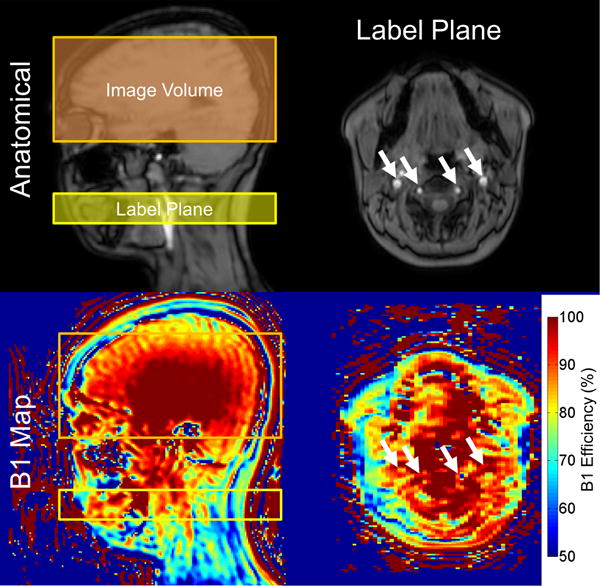
Top) Anatomic sagittal slice demonstrating the localization used in PCASL (left) and a transverse slice of the imaging plane (right). Internal carotid and vertebral arteries are denoted with white arrows. Bottom) Corresponding B1+ maps of the anatomical images. B1+ was significantly diminished at the intersection of the labeling plane and the cerebral feeding vessels compared to the imaging volume. Spatial heterogeneity in B1+ can lead to asymmetries in labeling efficiency that result in perceived asymmetries in CBF.