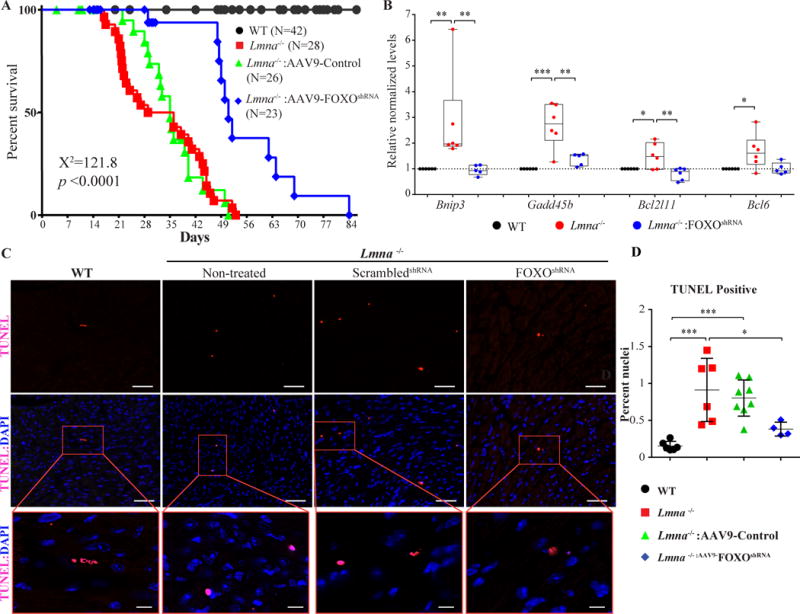
Figure 7. Phenotypic consequences of knock down of FOXO TFs in the Lmna−/− mouse hearts.

A. Kaplan-Meier survival plots of WT, Lmna−/− mice (not injected), Lmna−/− mice injected with AAV9-scrambledshRNA, or AAV9-Control (combined AAV9-scrambledshRNA and AAV9-Gfp) and Lmna−/− mice injected with the AAV9-FoxoshRNA constructs. The median survival time of Lmna−/− mice was increased from 29 days to 51 days upon administration of the AAV9-FoxoshRNA construct. The survival rates in the Lmna−/− mice injected with AAV9-Gfp or AAV9-scrambledshRNA constructs were similar to that in the (non-injected) Lmna−/− mice and are shown individually in Online FigureV. B. Transcript levels of selected markers of apoptosis, which are known to be FOXO TF targets, in the experimental groups. Transcript levels of BCL2 Interacting Protein 3 (Bnip3), Growth Arrest And DNA Damage Inducible Beta (Gadd45b), BCL2 Like 11 (Bcl2l11), and B-Cell CLL/Lymphoma 6 (Bcl6), quantified by RT-qPCR from mRNA extracted from 2-week old WT, Lmna−/− mice (not injected) and Lmna−/− mice injected with the AAV9-FoxoshRNA construct, were normalized in the treated group. C. Apoptosis assessments by TUNEL assay in the heart at 4 weeks of age. Representative TUNEL stained thin myocardial cross-section from 4-week old WT, Lmna−/− mice (not injected) and Lmna−/− mice injected either with AAV9-scrambledshRNA or the AAV9-FoxoshRNA constructs are shown. Upper panels show TUNEL staining in red and the middle panels show overlay of TUNEL (red) and DAPI-stained nuclei (blue) myocardial sections. The lower panels show magnification of selected areas displaying apoptotic cells. Bar is 50 μm in upper and middle panels, and 20 μm in the lower panels. Quantitative data of the TUNEL positive stained nuclei in the experimental groups are shown in panel D. The number of apoptotic cells was increased by 5.9 ± 1.13-fold in Lmna−/− mice (p=0.0008 vs WT); whereas the number in the AAV9-FoxoshRNA treated group was reduced by 58.2 ± 5.1 % (p=0.0247) compared to the non-treated Lmna−/− mice and tend to be decreased (p=0.071) compared to treated Lmna−/− mice. The number of apoptotic cells in the AAV9-FoxoshRNA treated Lmna−/− mice was not significant different from that in the WT mice (0.38 ± 0.05% vs. 0.15 ± 0.02%, respectively, p=0.55).
*: p<0.05, **: p<0.01, ***: p<0.001, ****: p<0.0001
