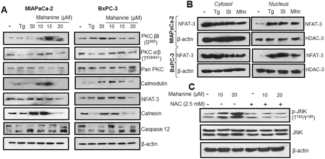
Figure 3.
Activation of Ca2+ signaling in mahanine treated-pancreatic carcinoma cells. (A) Molecular basis of activation of Ca2+ signaling was evaluated by western blot analysis in the cell lysate of MIAPaCa-2 and BxPC3 treated with increasing doses of mahanine (10, 15 and 20 µM) for 18 h. (B) Translocation of NFAT-3 from cytosol to nucleus in MIAPaCa-2 and BxPC-3 cells treated with 15 µM of mahanine (Mhn) for 18 hr. β-actin and HDAC-3 were used as loading control proteins for cytosolic and nucleus fractions respectively. (C) Enhanced phosphorylation of JNK in the MIAPaCa-2 cells treated with increasing doses (10 and 20 µM) of mahanine for 18 hr which was inhibited by pretreatment with NAC for 1 hr indicating the involvement ROS in this process.

