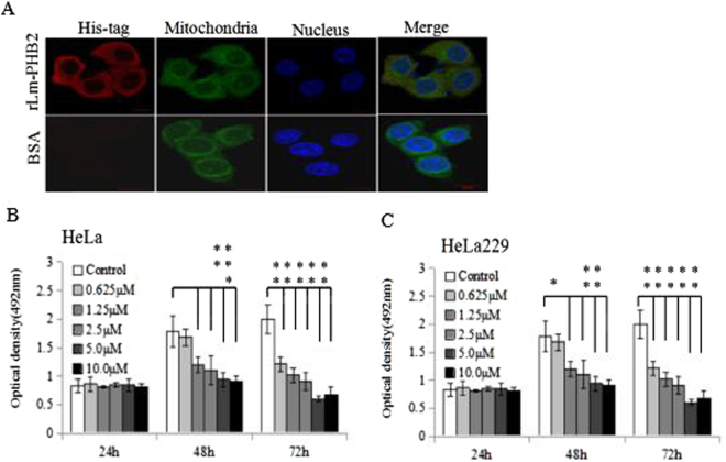
Figure 1.
Effect of purified rLm-PHB2 on the proliferation of human cervical cancer cells. (A) Confocal microscopic images of rLm-PHB2 protein entering into HeLa cells and localizing in cytoplasm. rLm-PHB2 culture 24 h were immunostained with His-tag antibody (Red). Mitochondria were stained with MitoTracker (Green), and nucleus were stained with Hoechst33258 (bule). Scale bar, 10 μm. (B,C) HeLa and HeLa 229 cells were treated with PBS or different concentrations of purified Lm-PHB2 (0.625 μM, 1.25 μM, 2.5 μM, 5.0 μM and 10.0 μM) at 37 °C for 24 h, 48 h or 72 h and cell viability was determined by the MTT assay. Data are the means ± SDs from three experiments, each carried out in triplicate. ‘*’ and ‘**’ indicate significantly different from control cells (not treated with Lm-PHB2) at the P < 0.05 and P < 0.01 levels, respectively.

