Figure 3.
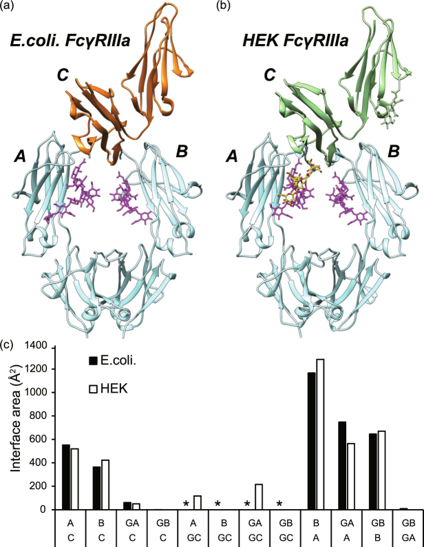
Comparison of structures of glycosylated and non-glycosylated FcγRIIIa in complex with IgG-Fc. (a) Crystal structure of Fc-FcγRIIIa complex. Recombinant Mut FcγRIIIa was expressed in Escherichia coli (non-glycosylated, left panel). (b) Previously reported crystal structure of Fc-FcγRIIIa complex (right panel, PDB ID: 3SGJ). Non-glycosylated FcγRIIIa, glycosylated FcγRIIIa, Fc, and the glycan attached to Fc are depicted in orange, light green, cyan, and magenta, respectively. The N-glycan molecule attached to Asn168 of FcγRIIIa is depicted in yellow sticks. The letters A, B, and C represent chains A and B of Fc, and FcγRIIIa, respectively. (c) Buried surface area (Å2) calculated by the PISA server51. GA, GB, GC indicate the N-glycan attached to Fc (chain A), Fc (chain B), and FcγRIIIa, respectively. The asterisks indicate that no interaction is possible for the non-glycosylated FcγRIIIa produced in E. coli.

