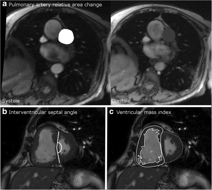
Fig. 1.
Diagram showing the methodology of calculation of the cardiac MRI metrics. Pulmonary artery relative area change (a) taken from cine images taken at the level of the pulmonary trunk, perpendicular to the main pulmonary artery; septal angle (b) taken as the angle made between the insertion points of the RV to the mid septum on the end-systolic image from the short axis stack; and (c) ventricular mass index taken by segmentation of the muscle mass of the left and right ventricle on the stack of images taken in the short axis plane

