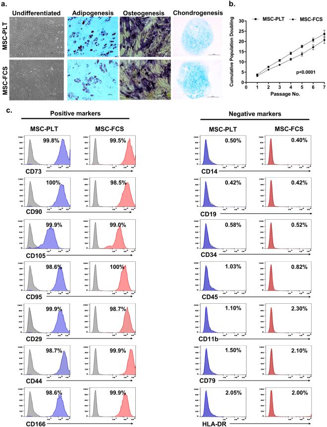
Figure 1.
Characteristics of MSC-PLT and MSC-FCS. (a) Representative images of MSC-PLT and MSC-FCS showing their morphology and ability to differentiate into adipocytes, osteoblasts and chondroblasts; Scale bars represent 100 or 200 μm; (b) Cumulative population doublings of MSC-PLT and MSC-FCS presented as the mean ± SEM of 13 independent samples; (c) Representative histograms of MSC-PLT and MSC-FCS phenotype showing comparable expression profile of the positive and negative markers.

