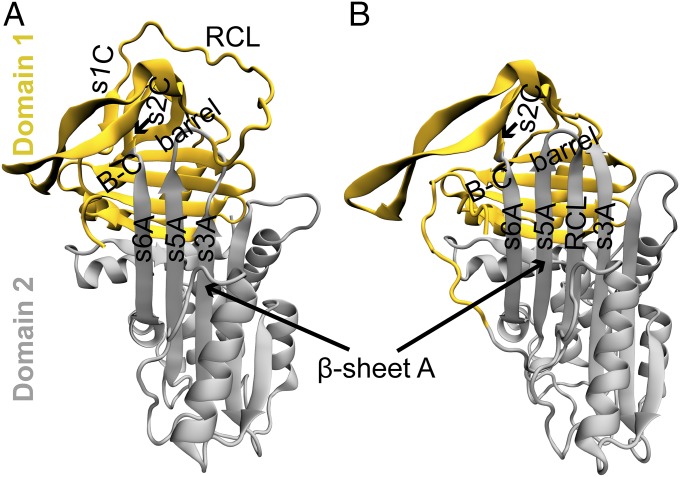
Fig. 2.
Domain definitions of the active and latent conformations of α1-AT. The structures of the (A) active and (B) latent conformations of α1-AT are colored according to their domains; α1-AT has two domains, according to the CATH database (23). Domains 1 and 2 are colored yellow and gray, respectively, in both conformations (see SI Appendix for definitions, and see SI Appendix, Fig. S1). Some of the key structural elements such as the RCL and strands s3A, s5A, and s6A are also marked. The RCL is in domain 1 in the active conformation, while it is a part of domain 2 in the latent conformation.

