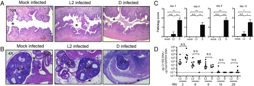
Fig. 1.
Infection with C. trachomatis serovar D induces immunopathology in the upper genital tract of female C57BL/6 mice. (A and B) Representative H&E-stained sections of uteri (A) and ovaries (B) from mock-infected, Ct L2-infected, and Ct D-infected mice at day 6 postinfection. Moderate-to-severe inflammation is observed only in the Ct D-infected mice, characterized by an influx of cells into the uterine lumen (* in A) and thickening and separation of the ovarian membrane from the ovary due to cellular infiltration (* in B). OD, oviduct; OV, ovary. (C) Upper genital tract pathology scores (0, none; 1, mild; 2, moderate; and 3, severe) for mock-infected, Ct L2-infected, and Ct D-infected mice. Day 3: mock vs. D, **P = 0.0020; L2 vs. D, **P = 0.0054. Day 6: mock vs. D, **P = 0.0083; L2 vs. D, **P = 0.0025. Day 9: mock vs. L2, **P = 0.0095; L2 vs. D, ***P = 0.0004. Day 15: mock vs. D, *P = 0.0257; L2 vs. D, *P = 0.0170. (D) Time course of bacterial burden in the upper genital tract of Ct L2- vs. Ct D-infected mice shows no significant differences between serovars. N.S., not significant. Day 3: P = 0.39. Day 6: P = 0.59. Day 9: P = 0.31. Day 15: P = 0.35. Day 29: P = 0.31.

