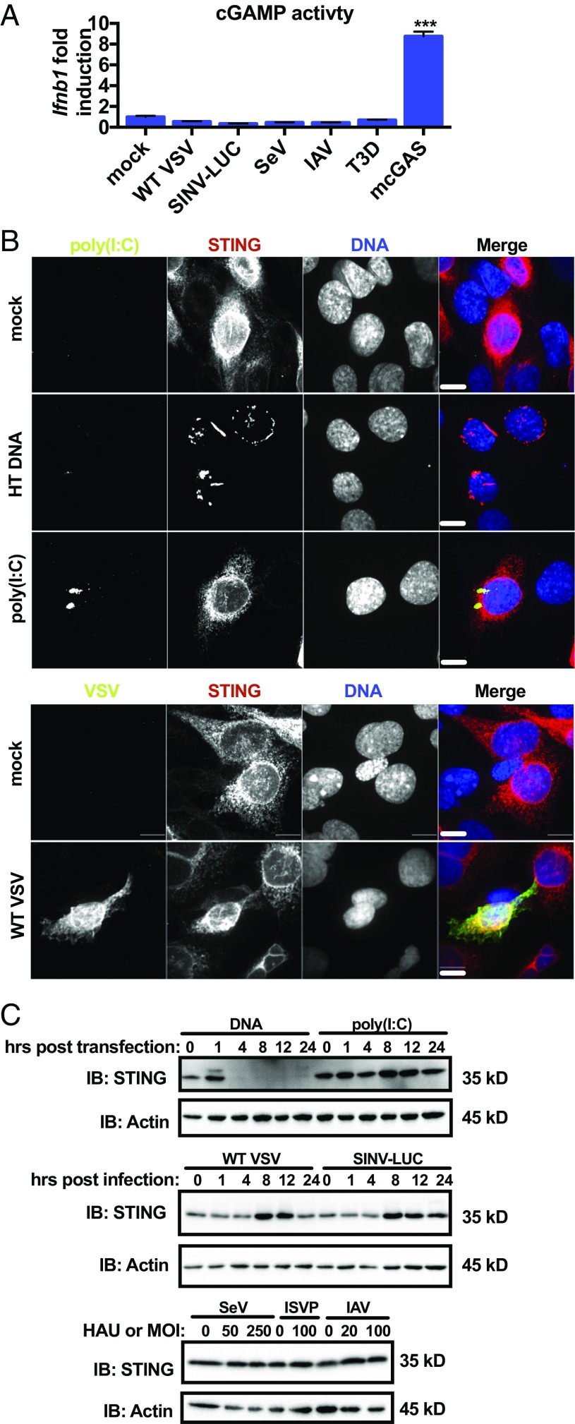
Fig. 2.
cGAS–STING pathway is not activated canonically during RNA virus infection. (A) WT MEFs were infected with VSV (MOI 10), SINV (MOI 10), SeV (250 HAU/mL), IAV (50 HAU/mL), or reovirus (MOI 100) for 18 h or transfected with a plasmid encoding mouse cGAS for 20 h. Virally infected or transfected cytoplasmic lysates were incubated with WT MEFs in the presence of reversible permeabilization buffer. Ifnb1 transcript was quantified by qRT-PCR. Data are displayed as fold induction of the indicated gene compared with uninfected. Data are represented as mean ± SEM. (B) WT MEFs expressing STING-HA were mock-transfected, transfected with herring testes DNA (5 μg/mL), poly(I:C) (2 μg/mL), mock-infected, or infected with VSV (MOI 1). MEFs transfected with DNA were fixed 1 h after transfection. Mock- and poly(I:C) transfected MEFs were fixed 4 h after transfection. VSV-infected and mock-infected MEFs were fixed 8 h after infection. MEFs were stained with antibodies detecting HA, dsRNA, or VSV M and stained to visualize nuclear DNA. Scale bar represents 10 μm. (C) WT MEFs were transfected with herring testes DNA (5 μg/mL), poly(I:C) (2 μg/mL), or infected with VSV (MOI 1) or SINV-LUC (MOI 1), SeV (0, 50, 250 HAU/mL), IAV (0, 20, 50 HAU/mL), or reovirus (MOI 100). SeV, IAV, and reovirus samples were collected 24 h postinfection. Lysates were separated by SDS/PAGE, and endogenous STING and actin were detected by western analysis. ***P < 0.0001 (Student’s t test).

