Fig. 3.
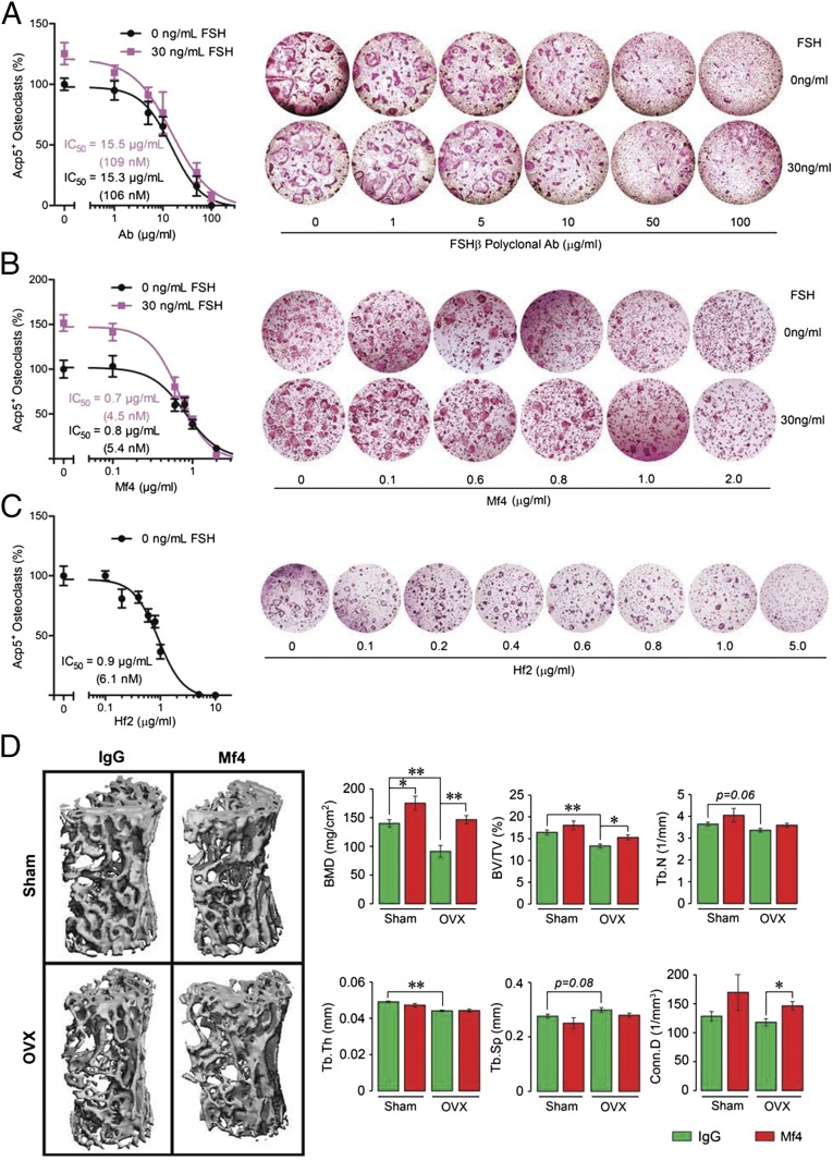
Monoclonal anti-epitope FSHβ antibody increases bone mass in ovariectomized mice. (A–C) To confirm FSH specificity of the monoclonal antibodies Mf4 and Hf2, Ficoll-purified hematopoietic stem cells were cultured with RANKL (30 ng/mL) and incubated with varying concentrations of the polyclonal FSH antibody (A), Mf4 (raised against CLVYKDPARPNTQKV) (B), or Hf2 (C) for 5 d in the presence or absence of mouse Fsh (30 ng/mL) (n = 8 wells per group). Cells stained using a kit for tartrate-resistant acid phosphatase (Acp5) were counted. Representative culture wells are shown, together with mean percent osteoclast number (±SEM). Note that the increase in osteoclastogenesis noted with 30 ng/mL FSH (and zero-dose antibody) was abolished progressively with increasing antibody concentrations. IC50s for the polyclonal FSH antibody, Mf4 and Hf2 are shown. (D) Mice were ovariectomized or sham-operated and injected with Mf4 or mouse IgG (100 µg/d) for 4 wk while on normal chow (n = 5 mice per group). The vertebral column was dissected and processed for micro-CT measurements. Representative micro-CT images and structural parameters, namely BMD, BV/TV, Tb.Th, Tb.N, Tb.Sp, and/or Conn.D, are shown. Statistics: mean ± SEM; two-tailed Student’s t test, corrected for multiple comparisons; *P ≤ 0.05, **P ≤ 0.01, or as shown.

