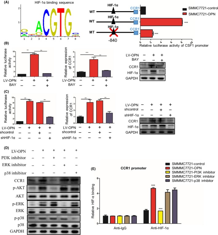
Figure 5.

Osteopontin (OPN) upregulates CCR1 expression via PI3K/Akt/HIF‐1α pathway. A, SMMC7721 cells with or without lentivirus (LV)‐OPN were transiently transfected with a wild‐type CCR1 promoter‐dependent luciferase construct or a mutant construct wherein HIF‐1α binding sites were mutated (WT: GGCACGTGCAG; MT: GGCATAGACAG). Luciferase activity was detected 24 h after LV‐OPN transfection. B, SMMC7721‐OPN cells were treated by HIF‐1α inhibitor BAY 87‐2243. The promoter activity and expression of CCR1 were measured by luciferase reporter assay, quantitative PCR (qPCR) and western blot, respectively. C, SMMC7721‐OPN cells were transfected with HIF‐1α shRNA or control shRNA. The promoter activity and expression of CCR1 were measured by luciferase reporter assay, qPCR and western blot, respectively. D, SMMC7721‐OPN were treated by PI3K, ERK or p38 inhibitors. The expression of CCR1, phosphorylated and total AKT, ERK and p38 were analyzed by western blot. E, ChIP‐qPCR of HIF‐1α on the CCR1 promoter in the presence of LV‐OPN and PI3K inhibitor. Data are expressed as means ± SEM and compared using Student's t‐test (*P < .05, **P < .01 and ***P < .001)
