Figure 1.

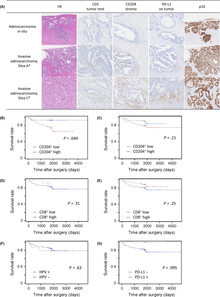
(A) Representative images of CD3+, CD204+, and programmed cell death 1 ligand‐1 (PD‐L1) expression and p16 immunohistochemistry according to tumor histology based on the Silva classification of invasion patterns. Histology (H&E staining) is categorized into adenocarcinoma in situ and Silva patterns A (well‐demarcated glands without desmoplastic reaction or lymphovascular invasion) and C (extensive destructive stromal invasion). Corresponding immunohistochemistry images are shown. (B‐G) Kaplan–Meier survival curves for 5‐year disease‐free survival in patients with cervical adenocarcinoma. Patients were dichotomized by median density of tumor‐infiltrating lymphocytes (TILs) or tumor‐associated macrophages (TAMs). Stromal infiltration of CD204+ TAMs (B) and CD8+ TILs (D), infiltration of the tumor nest by CD204+ TAMs (C) and CD8+ TILs (E), human papillomavirus (HPV) status (F), and expression of PD‐L1 on tumor cells (G). Data were statistically analyzed by log–rank test
